"Can spay be done together
with the removal of sternal abscess?" the young man who
brought in his old dog to the surgery as scheduled, on this
bright Sunday May 18, 2008 morning asked.
 |
|
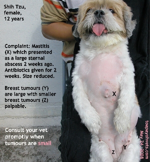
"Sternal abscess" ---
actually mastitis, now reduced after 2 weeks of
antibiotics |
"Yes, it can be done" I explained. "It is not good for the old
dog as the anaesthetic time will be longer. The chances of the
old dog dying on the operating table increases as the
anaesthesia is prolonged. The spay itself takes around one
hour."
"I thought it takes 30
minutes to spay a dog?" the young man must have done his home
work and research.
"Yes, in some cases" I said but did not elaborate that his dog
was a bit on the fat side and spay surgery would take longer.
"The actual surgery can be as fast as 30 minutes from skin
incision to last stitch of the skin if the vet can hook up the
uterine horn at the first try.
"However, if you include the pre-operation shaving, scrubbing
and anaesthetic gas given to get the dog to sleep, the whole
spay takes more than 1 hour. In removing the sternal abscess,
I need to pull skin from nearby areas, so it is not a simple
stitching of just the wound left from removing the sternal
abscess. This takes a lot of time, around 30 minutes or more.
"
He nodded his head. Do one surgery at a time to avoid
anaesthetic complications. Spay today. Then 2 weeks later,
remove the mammary tumour and sternal abscess.
So, I started to time this spay surgery commenced at 10 am.
The patient was not slim and had difficulty breathing, being a
flat-faced Shih Tzu.
Procedure:
The dog was clipped at the preparation room. Then she was
brought to operating room and given 8% gas to knock her down
using a face mask. She struggled for 2 minutes and was asleep.
This took around 15 minutes. She was then intubated with a
breathing tube to connect her lungs to the gas machine, using
a maintenance dose 2% gas until the last 3 stitches when the
gas was reduced to 0%. Dog woke up within 2 minutes at end of
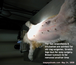
anaesthesia. The four legs were stretched out tautly as I find
this method enabled me to hook up the uterine horn much
easier.
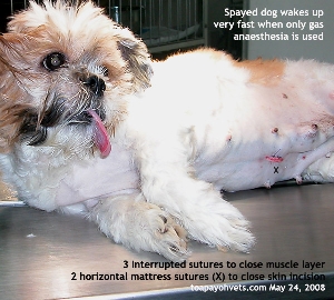
 |
|
Gas anaesthesia is
best and safe for old dogs. Do intubate all the time
although gas mask can be used |
 |
|
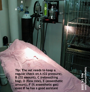
The vet must check
the anaesthetic settings systematically to ensure a
smooth anaesthetic process |
Surgery:
Incision to last stitch was 40 minutes actually.
I incised 1 cm from the umbilicus, making a 1-cm cut. The
linea alba was identified after snipping off some subcutaneous
fat. There was some bleeding but this was not serious.
I inserted the spay hook to fish out the left uterine horn.
I slanted the hook 45 degrees from the horizon, put it into
the abdomen to the right and downwards in the direction of the
bladder. The hook skimmed over the surface the liver lobes.
Then I rotated the hook 90 degrees and pulled it out of the
skin incision, hopefully with the left uterine horn. In slim
female dogs, this was not a problem. But in this dog, I tried
8 times. Omental fat kept appearing in the hook. This was not
good as the minutes passed quickly. In such cases, I switch to
hooking the right uterine horn. One loop of pink intestine
kept coming out in the hook. What to do? Persevered.
If the dog was slim, it would normally be easy for me to hook
the left uterine horn with the left ovary after 1 or 2 tries.
Fortunately, I caught the right uterine horn on the 3rd try. What
a relief you would imagine.
But there was so much fat surrounding the right ovary. I could
not fish it out with the right uterine horn. "Release the
string's tension on front legs," I asked my assistant to
loosen the strings."
Still, the ovary could not be hooked out. I knew I had to
extend the skin and linea alba incision 0.5 cm cranially. This
bigger incision was sufficient. I pulled out the right fat-
enclosed ovary. I felt for the taut ovarian ligament with my
left forefinger. A very tight ligament. The dog moved as
the ovarian ligament was pulled.
"Increase gas to 5% for 30 seconds and then go back to 2%," I
said to the assistant. This happened when the dog was just
slightly under surgical anaesthesia and had not felt any pain
till the ovarian ligament was pulled. Previously I used to
pull this the ligament broke from its attachment. Nowadays, I
used the scalpel to break it and continued ligating the
ovarian stump.
I ligate the stump 2 times after placing 3 artery forceps
clamps on the ovarian tissues cranial to the ovary. Now, the
ovary could not be seen as it was enveloped inside a thick
clump of fat. I had to estimate its position.
After ligation and incision of the ovarian fat, I lifted up the
right uterine horn. The assistant had loosened the tension of
the front legs. In some cases, I asked the assistant to put
his hands under the shoulders and elevated the dog so that I
could access the uterine horn easier. In this case, on pulling
the right uterine horn, I could see the uterine body and the
left uterine horn arising from there.
The same process of getting the ovarian ligament was repeated.
Then the uterine body was clamped using the 3-forcep technique
as for the ovarian tissue. 2 ligations were used.
3/0 absorbable sutures did not feel strong enough compared to
2/0 but was used in this dog. I closed the muscle layer with 3
simple interrupted sutures placed a good 5 mm away from the
muscle edge. If you place it too close, the suture might
break down and you would get a hernia.
 |
|
Reduce gas to 0%
before after closing the muscle layer. Dog wakes up as
the last skin stitch is placed |
It would be best not to
use continuous sutures to close the muscle layer unless you
are very confident of your suture placement. Otherwise one too
close to the muscle edge stitch may burst open. The whole
stitching breaks down and there would be a lump. Also, I do
not use continuous subcutaneous suture as advised in some
veterinary surgery books. They cause more irritation and may
break down. I used 2 horizontal mattress sutures to close up
the skin incision. One packet of suture was fully used in this
case. In bigger sized dogs, 2 packets may be needed.
Around 2 cotton swabs would be used as there was little
bleeding.
 |
|
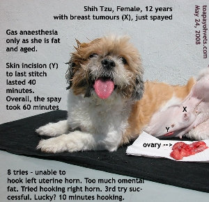
12-year-old Shih
Tzu 7.7 kg, 38.4 deg before spay. Large mammary
tumours (X). First skin incision to last skin stitch
took 40 minutes in this case as the first time hooking
did not fish out the uterine horn.
|
Carprofen a
non-steroidal anti-inflammatory injection 0.5 ml was given to
prevent pain and swelling. Antibiotics given. The dog went
home in the evening after sufficient rest at the surgery. Do
not send the dog home immediately even if the owner wanted to
do so as the dog needed time to be stable after anaesthesia.
NEXT STAGE
Spay was advised first to remove the female hormone production
by the ovaries. Once deprived of the hormones, it was hoped
that the breast tumours would not grow so aggressively. 2
weeks later, the breast tumour and the sternal abscess would
be removed, hopefully without any anaesthetic complications
and death.
CONCLUSION
Can this dog survive the anaesthesia the second time?
Nobody can guarantee survival under general anaesthesia. In
any case, never attempt to spay and remove breast tumours in
one surgery as the vet prolongs anaesthesia time. Every second
that the dog is under anaesthesia, his or her heart may fail.
So, it is best to do one surgery at a time to minimise the
risk.
 UPDATE
AS AT FEB 11, 2010 UPDATE
AS AT FEB 11, 2010
The owner did not return to Toa Payoh Vets for surgery nor
follow up. Spaying the dog when she was young would be ideal
as breast tumours seldom occur in spayed female dogs. This is
not to say that spayed female dogs don't get breast tumours
but the probability of them getting such tumours are much
lower. |